
Eight fields. One standard of proof.
The catalog filed the way laboratories actually work — by receptor family and pathway. Each area lists what is studied, the models commonly used, and the compounds we stock for it.
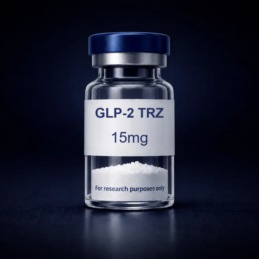 01GLP & metabolic5 compounds · from $80.00
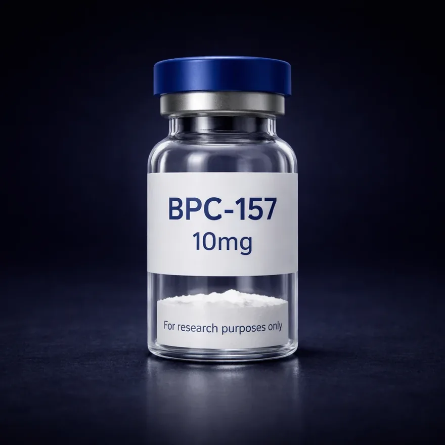 02Repair & recovery4 compounds · from $55.00
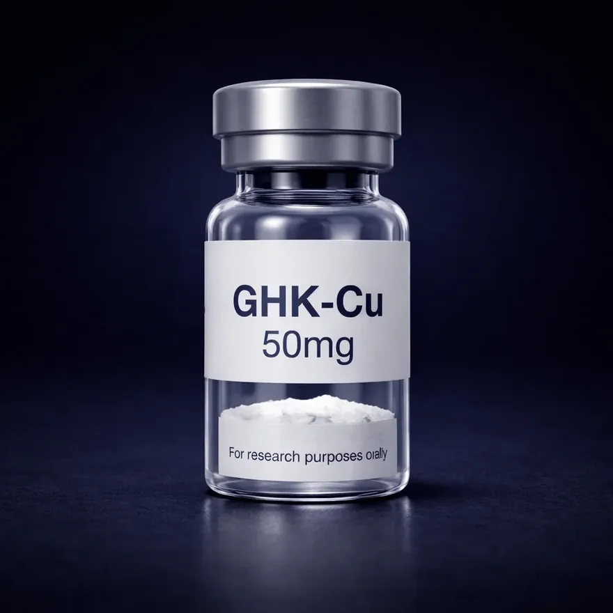 03Skin & cosmetic4 compounds · from $35.00
 04Neuro & cognitive3 compounds · from $45.00
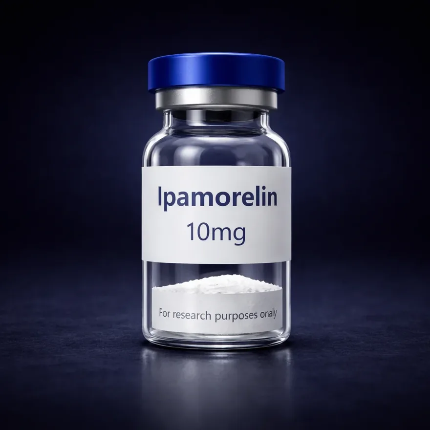 05GH secretagogues4 compounds · from $70.00
 06Mitochondrial3 compounds · from $55.00
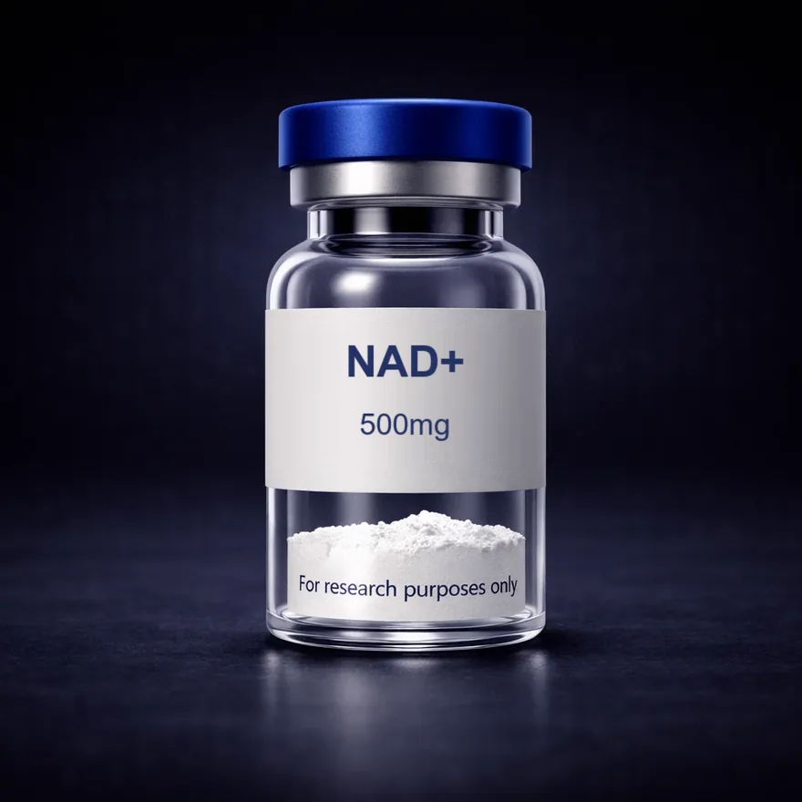 07Vitamins & cofactors4 compounds · from $40.00
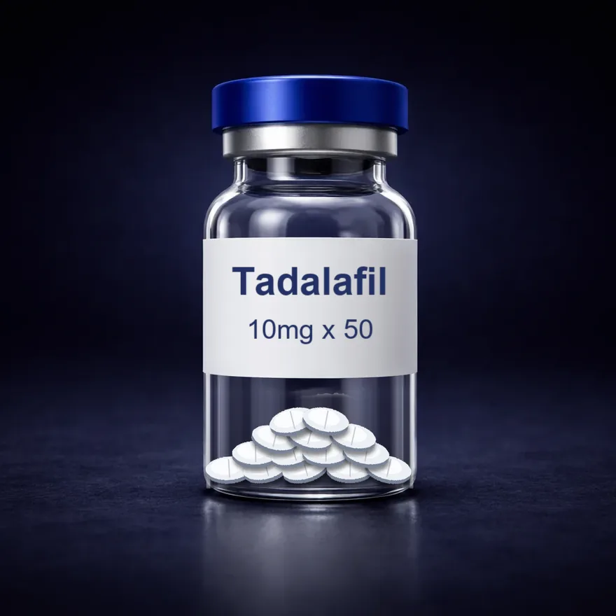 08Small molecules1 compound · from $65.00
 How to read this page. Areas describe the research each compound is used in — not effects in people or animals. RenuPep products are for in-vitro laboratory research only.
GLP & metabolic
Incretin and metabolic-signalling research.
This area covers the receptor systems behind nutrient-response signalling — the GLP-1, GIP, glucagon and amylin receptors. Compounds range from single-receptor agonists to dual and triple agonists, which makes the group useful for comparative work on receptor selectivity.
Laboratories study how receptor activation is carried through cAMP and other second messengers, how sequence modifications change stability in solution, and how multi-receptor sequences compare with single-receptor ones in the same assay.
What laboratories study
- Receptor binding and selectivity assays
- cAMP and second-messenger signalling
- Comparative incretin-analogue studies
- Solution stability and degradation profiling
Common models & methods
- Receptor-expressing HEK293 cell lines
- Pancreatic beta-cell lines (INS-1, MIN6)
- cAMP accumulation assays
- HPLC stability profiling
- GLP-1R
- The glucagon-like peptide-1 receptor, a class B G-protein-coupled receptor.
- GIP
- Glucose-dependent insulinotropic polypeptide, an incretin hormone with its own receptor, GIPR.
- Amylin analogue
- A synthetic sequence modelled on amylin, a peptide co-secreted with insulin, used in receptor studies.
Repair & recovery
Cellular-signalling and tissue-response research.
Peptides in this group are studied for how short sequences interact with growth-factor pathways, the cytoskeleton and the extracellular matrix.
Typical work uses cell-migration and scratch-closure assays, endothelial tube formation and cytokine panels, all under controlled in-vitro conditions.
What laboratories study
- Cell migration and scratch-closure assays
- Growth-factor and angiogenic signalling
- Extracellular-matrix expression studies
- Cytokine and inflammatory-marker models
Common models & methods
- Fibroblast and endothelial cell culture
- Scratch-closure migration assays
- Tube-formation assays
- Cytokine and ELISA marker panels
- Thymosin β4
- A 43-amino-acid actin-sequestering peptide. TB-500 is a synthetic fragment used in research.
- Pentadecapeptide
- A peptide fifteen amino acids long, such as BPC-157.
- Actin regulation
- How cells build and break down actin filaments — central to cell-migration studies.
Skin & cosmetic
Dermal-signalling and pigment-pathway research.
Two families sit here: copper-peptide complexes and melanocortin-receptor analogues, alongside multi-compound research blends.
Laboratory work looks at how these compounds bind, which cascades they engage, and how they behave in fibroblast and melanocyte culture models.
What laboratories study
- Melanocortin receptor (MC1R–MC5R) binding
- Fibroblast collagen-expression assays
- Copper-complex stability and chelation
- Melanogenesis pathway analysis in culture
Common models & methods
- Dermal fibroblast culture
- Melanocyte and B16 cell lines
- Collagen and elastin expression assays
- Copper-binding and chelation analysis
- GHK-Cu
- The tripeptide glycyl-L-histidyl-L-lysine complexed with copper(II).
- Melanocortin receptors
- A family of five G-protein-coupled receptors, MC1R to MC5R, that bind α-MSH and its analogues.
- Melanogenesis
- The biochemical pathway by which melanocytes synthesise melanin.
Neuro & cognitive
Neuropeptide and neurochemical-signalling research.
These sequences are modelled on endogenous regulatory fragments — ACTH(4–7) in the case of Semax, and tuftsin in the case of Selank, each extended with a Pro-Gly-Pro tail.
In-vitro studies examine neurotransmitter-system signalling, neurotrophic-factor expression, and how short peptides resist or undergo enzymatic degradation.
What laboratories study
- Neurotrophic factor expression assays
- Monoamine and GABAergic signalling
- Enzymatic stability of short peptides
- Receptor binding in neuronal cell lines
Common models & methods
- Neuronal cell lines (SH-SY5Y, PC12)
- Neurotrophin expression assays
- Enzymatic degradation studies
- Receptor-binding studies
- ACTH(4–7)
- A four-residue fragment of adrenocorticotropic hormone. Semax adds a Pro-Gly-Pro tail to it.
- Tuftsin
- A four-amino-acid peptide. Selank extends its sequence with a Pro-Gly-Pro tail.
- BDNF
- Brain-derived neurotrophic factor — a common readout in neurotrophin expression assays.
GH secretagogues
Growth-hormone-axis research.
The group spans GHRH analogues, selective ghrelin-receptor agonists and IGF-pathway compounds — the full signalling axis in one place.
Research examines receptor selectivity, secretion models in pituitary cell culture, and how structural modifications change a sequence’s stability.
What laboratories study
- GHRH and GHS-R1a receptor binding
- Pulsatile secretion in pituitary culture
- IGF-1 receptor kinase signalling
- Structural modification and stability
Common models & methods
- Pituitary cell culture
- GHRH-R and GHS-R1a binding assays
- IGF-1R phosphorylation assays
- Structure–stability comparisons
- GHRH
- Growth-hormone-releasing hormone. CJC-1295 and Tesamorelin are synthetic analogues.
- GHS-R1a
- The ghrelin receptor, targeted by selective secretagogues such as Ipamorelin.
- IGF-1 LR3
- A long-chain IGF-1 analogue with an arginine at position 3 and a 13-residue N-terminal extension.
Mitochondrial
Mitochondrial and cellular-energy research.
This area brings together a mitochondrial-derived peptide, a cardiolipin-binding peptide and a small-molecule ERR agonist.
Work here examines mitochondrial membrane integrity, oxidative stress and the signalling that links a cell’s energy status to metabolic regulation, typically in isolated mitochondria and cultured cell lines.
What laboratories study
- Membrane potential and respiration assays
- Reactive oxygen species measurement
- AMPK and energy-sensing pathways
- Cardiolipin and membrane interaction
Common models & methods
- Isolated mitochondria preparations
- Extracellular-flux respiration assays
- ROS and membrane-potential dyes
- AMPK pathway analysis
- Cardiolipin
- A phospholipid of the inner mitochondrial membrane. 2S-31 binds to it.
- MOTS-c
- A 16-amino-acid peptide encoded within mitochondrial DNA.
- ERR
- Estrogen-related receptors — orphan nuclear receptors involved in energy-metabolism gene expression.
Vitamins & cofactors
Coenzyme, redox and micronutrient research.
This group covers the cofactors and micronutrients that cellular chemistry depends on: NAD+, the redox coenzyme; glutathione, the principal intracellular thiol antioxidant; and vitamin B-12 formulations.
Laboratories use them to study redox balance, methylation and one-carbon metabolism, and how energy-metabolism enzymes respond when cofactor supply changes in culture.
What laboratories study
- Redox balance and ROS scavenging assays
- NAD+-dependent enzyme activity
- Methylation and one-carbon metabolism
- Solution stability and degradation profiling
Common models & methods
- NAD+/NADH ratio assays
- GSH/GSSG redox assays
- Sirtuin and PARP activity assays
- Methylation pathway analysis
- NAD+
- Nicotinamide adenine dinucleotide in its oxidised form, a coenzyme central to cellular redox reactions.
- Glutathione
- The tripeptide γ-glutamyl-cysteinyl-glycine, the most abundant intracellular thiol antioxidant.
- Cyanocobalamin
- A stable synthetic form of vitamin B-12, a cofactor for methionine synthase and methylmalonyl-CoA mutase.
Small molecules
Enzyme-inhibitor and second-messenger research.
Small molecules are low-molecular-weight, non-peptide compounds. Tadalafil, stocked here, is a selective inhibitor of phosphodiesterase type 5 (PDE5).
Laboratory work uses PDE5 inhibitors to study cGMP signalling — how the second messenger is made and broken down, and how that balance behaves in smooth-muscle and endothelial cell models.
What laboratories study
- PDE isoform selectivity assays
- cGMP second-messenger signalling
- Smooth-muscle and endothelial cell models
- Solubility and stability profiling
Common models & methods
- PDE enzyme inhibition assays
- cGMP quantification (ELISA)
- Smooth-muscle cell culture
- Endothelial nitric-oxide signalling models
- PDE5
- Phosphodiesterase type 5, an enzyme that breaks down cyclic GMP.
- cGMP
- Cyclic guanosine monophosphate, an intracellular second messenger.
- IC50
- The inhibitor concentration that halves enzyme activity in a given assay.
Not sure where a compound sits?
Ask support — they can tell you how a compound is classified, what its certificate covers, and what is in stock today.